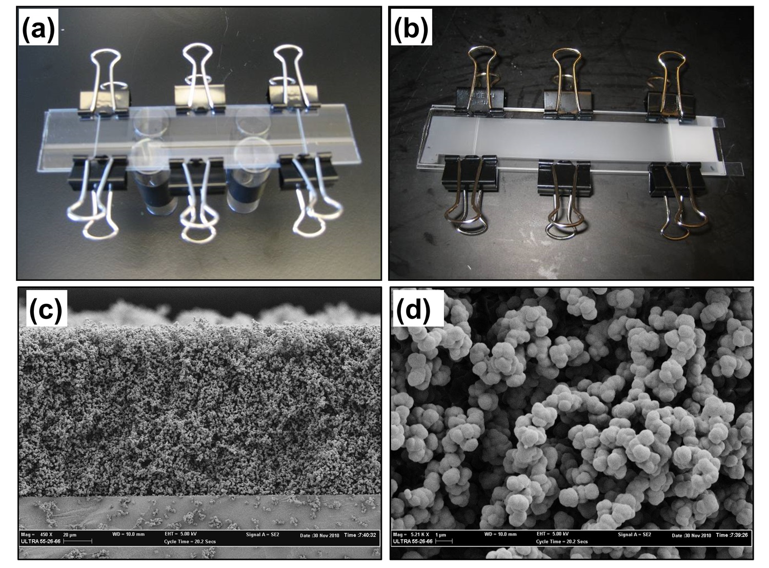
Figure 5-8. Mold consisting of two glass plates separated by Teflon strips located along the long side used for the preparation of monolithic layer (A), the mold containing the white monolithic layer (B), scanning electron microscope image of the cross section of the monolith (C), and detailed view of the morphology of the monolithic layer (D).
*Click on image for full-size .jpg*
<< Back to Chapter 5 <<